The Acoustic Physics and Cellular Dynamics of Ultrasonic Cavitation in Aesthetic Body Remodeling

The clinical evolution of non-invasive body contouring has transformed aesthetic medicine, providing alternatives to surgical interventions such as tumescent liposuction and mechanical panniculectomy. For decades, the removal of localized subcutaneous adipose deposits required invasive mechanical disruption, general anesthesia, surgical incisions, and prolonged recovery periods accompanied by risks of hematoma, infection, and contour irregularities. As patient demand shifted toward outpatient procedures characterized by minimal downtime, medical physicists and biomedical engineers explored the direct therapeutic application of acoustic energy. Ultrasonic waves, historically confined to diagnostic sonography and high-energy surgical cutting, were re-engineered to deliver focused mechanical forces directly into biological tissue.
Within modern clinical protocols, acoustic field manipulation allows practitioners to target subcutaneous fat layers while preserving surrounding structures, including vascular networks, peripheral nerve bundles, and the overlying epidermal barrier. Deploying a professional cavitation machine relies on the precise calibration of acoustic frequencies, transducer geometries, and energy densities to induce stable and transient mechanical oscillations within interstitial fluids. Understanding the therapeutic efficacy and physical boundaries of this technology requires an examination of the acoustic physics of cavitation, the histology of adipose tissue, the metabolic pathways responsible for lipid clearance, the integration of complementary radiofrequency energy, and the clinical safety frameworks governing acoustic body remodeling.
Fundamental Principles of Acoustic Wave Propagation and Wavefront Mechanics
To analyze how acoustic energy alters biological structures, one must examine the physical properties of sound waves as they propagate through heterogeneous organic matter. Sound travels through physical media as a longitudinal mechanical wave characterized by alternating cycles of spatial compression and rarefaction. During the compression phase, molecules within the medium are forced together under elevated localized pressure; conversely, during the rarefaction phase, molecules are pulled apart, establishing a transient negative pressure state.
The velocity, attenuation, and absorption of these acoustic waves are governed by the acoustic impedance of the specific tissue layer. Acoustic impedance represents the product of a medium’s physical density and the speed of sound within that medium. When an acoustic beam travels through human tissue, boundaries between distinct histological layers—such as the transition from the dermis to subcutaneous adipose tissue, or from adipose tissue to deep muscular fascia—present impedance mismatches.
These boundary interfaces cause a portion of the wave energy to reflect while the remaining fraction refracts and continues deeper into the tissue envelope. Because adipose tissue possesses low acoustic impedance relative to dense muscle or bone, acoustic energy passes through subcutaneous fat with minimal attenuation, provided the operating frequency is selected to prevent early absorption in the superficial dermal strata.
See also: How to Use a Sauna Heater: Temperature, Timing, and Etiquette for Beginners
The Physical Mechanics of Stable and Transient Cavitation
Cavitation describes the formation, growth, and subsequent dynamic behavior of microscopic vapor-filled cavities or micro-bubbles within a liquid medium subjected to rapid pressure fluctuations. In biological systems, the targeted liquid is the interstitial fluid surrounding individual adipocytes within the subcutaneous matrix.
When acoustic waves propagate through this fluid at sufficient acoustic pressures, the tensile stress generated during the negative-pressure rarefaction phase exceeds the local intermolecular tensile strength of the liquid. This mechanical rupture creates microscopic cavities. The behavior of these micro-bubbles is divided into two distinct physical classes:
Stable Acoustic Cavitation
Under lower acoustic pressure regimes, micro-bubbles oscillate symmetrically around an equilibrium radius over numerous continuous acoustic cycles. As the surrounding pressure fluctuates, the bubble expands during rarefaction and contracts during compression without experiencing violent structural collapse. This periodic oscillation generates localized fluid circulation known as micro-streaming. Micro-streaming exerts sustained shear stresses against adjacent biological membranes, increasing cell membrane permeability without necessarily causing immediate structural rupture.
Transient or Inertial Cavitation
When acoustic power increases or frequencies are calibrated to specific resonance windows, the micro-bubbles undergo rapid, unstable growth over a small number of cycles. As the bubble expands, it absorbs energy from the acoustic field until it reaches an unstable critical radius. During the subsequent compression phase, the surrounding liquid rushes inward with extreme velocity, causing the bubble to violently implode.
The collapse of an inertial cavitation bubble produces localized physical phenomena, including shockwaves characterized by pressures exceeding hundreds of atmospheres and localized micro-jets of liquid traveling at velocities of hundreds of meters per second. When these micro-jets strike adjacent adipocyte membranes, the localized shear stresses overcome the mechanical tensile strength of the phospholipid bilayer, forming microscopic pores or completely rupturing the cellular envelope.
Resonance Tuning and Transducer Frequency Selection
The frequency of the acoustic wave dictates both the penetration depth and the size of the cavitation bubbles generated within the tissue. In aesthetic cavitation systems, operating frequencies are typically engineered within the low-frequency ultrasonic band, primarily between twenty-eight and forty kilohertz.
Lower frequencies produce larger resonant bubble diameters. A bubble resonating within a forty-kilohertz acoustic field expands to a substantially greater physical volume before undergoing inertial collapse than a bubble subjected to megahertz-frequency diagnostic ultrasound. The implosion of these larger bubbles releases the kinetic energy required to mechanically disrupt fibrous connective septa and resilient adipocyte walls.
Furthermore, low-frequency ultrasound experiences low absorption rates in superficial water-rich tissue, allowing the acoustic wave to bypass the epidermis and papillary dermis without generating excessive cutaneous heat. The energy concentrates within the subcutaneous layer, where the acoustic beam can be shaped or focused through concave piezoelectric transducer faces to achieve peak pressure thresholds at calibrated anatomical depths between one and three centimeters beneath the skin surface.
Adipose Histology and the Mechanisms of Adipocyte Membrane Disruption
Subcutaneous adipose tissue is organized into lobules separated by fibrous septa composed of Type I and Type III collagen, containing blood vessels, lymphatic capillaries, and unmyelinated nerve fibers. The functional parenchymal cells—mature white adipocytes—are spherical structures ranging from fifty to one hundred and fifty micrometers in diameter. Unlike other human cells that maintain complex cytoplasmic internal architecture, a mature adipocyte is largely occupied by a single, unilocular lipid droplet composed of neutral triglycerides, which displaces the cytoplasm and nucleus to the periphery against the plasma membrane.
Because the interior of the adipocyte consists of non-polar triglycerides, it behaves differently from the surrounding polar interstitial fluid under acoustic stress. When inertial cavitation occurs in the extracellular fluid immediately adjacent to the adipocyte, the resulting directional micro-jets and high-pressure shockwaves exert mechanical forces on the thin peripheral membrane.
Because the intracellular lipid droplet lacks the fluidity to deform rapidly and absorb high-velocity mechanical shocks, a high shear gradient forms across the phospholipid membrane. This physical force causes structural failure of the membrane, releasing stored triglycerides into the interstitial fluid matrix.
Metabolic Pathways of Lipid Clearance and Systemic Elimination
The mechanical destruction of adipocytes releases intracellular contents—comprising triglycerides, free fatty acids, glycerol, and cellular debris—into the extracellular compartment. Because insoluble lipids cannot enter the systemic bloodstream directly without risking fat embolism, the clearance of these mobilized compounds relies on natural lymphatic and hepatic pathways.
Glycerol is a water-soluble molecule that diffuses directly through vascular capillary fenestrations into the bloodstream. Once in the systemic circulation, it is transported to the liver and kidneys, where it is either converted into glucose via gluconeogenesis or channeled into the glycolytic cycle to generate adenosine triphosphate.
Triglycerides and long-chain fatty acids, by contrast, are hydrophobic and cannot enter standard blood capillaries directly. Instead, they are taken up by terminal lymphatic capillaries present within the adipose tissue septa. Specialized immune cells, primarily resident macrophages, migrate to the site of cellular breakdown, engulfing free lipid droplets and fragmented membrane components through phagocytosis.
The lymphatic fluid transports these lipids through regional lymph nodes and along major lymphatic trunks, eventually discharging them into the venous circulation via the thoracic duct. Once in the bloodstream, the lipids bind to serum albumin and are transported to the liver. Hepatic tissue processes these mobilized fatty acids through beta-oxidation pathways or incorporates them into endogenous lipoprotein complexes, such as very-low-density lipoproteins, for distribution to skeletal muscle and organs for metabolic energy expenditure. Consequently, the rate of physical lipid clearance depends on the functional efficiency of the patient’s lymphatic system and basal hepatic metabolic capacity.
Multi-Modal Synergies: Integrating Radiofrequency and Vacuum Drainage
While low-frequency acoustic cavitation addresses localized subcutaneous fat deposits, standalone cavitation does not directly treat dermal laxity or accelerate lymphatic drainage. Advanced treatment platforms overcome these limitations by combining ultrasonic cavitation with complementary physical modalities, primarily multi-polar radiofrequency and mechanical vacuum therapy.
Radiofrequency energy operates through alternating electrical currents that encounter natural impedance within biological tissue, generating volumetric heat in accordance with Joule’s law. While ultrasound disrupts the deep fat layer mechanically, radiofrequency delivers thermal energy to the upper reticular dermis, heating structural collagen fibers to temperatures between forty and forty-three degrees Celsius.
This thermal elevation causes immediate denaturation and triple-helix contraction of existing collagen strands, followed by the stimulation of dermal fibroblasts to synthesize new collagen and elastin over subsequent months. Combining these modalities ensures that as subcutaneous fat volume decreases, the overlying cutaneous envelope contracts, preventing loose or sagging skin contours.
Pneumatic vacuum integration serves a mechanical drainage purpose. By applying rhythmic, controlled negative pressure to the treated tissue fold, the vacuum handpiece stretches the fibrous connective tissue, increases micro-vascular perfusion, and mechanically drives mobilized interstitial fluids toward primary regional lymph nodes. This active lymphatic clearance reduces post-treatment edema, accelerates the removal of emulsified triglycerides, and optimizes acoustic wave transmission by keeping target tissues uniformly hydrated.
Operational Parameters, Dosimetry, and Clinical Governance
Safe and effective application of ultrasonic cavitation requires adherence to calibrated dosimetry and operational protocols. Delivering insufficient acoustic energy fails to cross the mechanical threshold required to trigger inertial cavitation, yielding negligible clinical results. Conversely, delivering excessive energy or holding the transducer stationary over a single point can cause acoustic energy to reflect off deep periosteal bone layers, generating painful standing waves and localized periosteal heating.
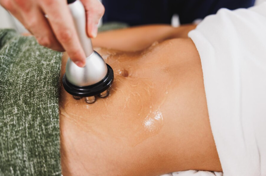
Practitioners follow continuous dynamic movement patterns, keeping the transducer head in slow, constant motion across the treatment zone while maintaining uniform contact through high-viscosity ultrasound coupling gels. The coupling medium is essential: because air possesses high acoustic impedance relative to biological tissue, even microscopic air gaps between the transducer face and the skin reflect nearly one hundred percent of the emitted acoustic energy, preventing penetration and risking thermal damage to the transducer crystal.
Clinical evaluations must also screen for physiological contraindications. Because mobilized triglycerides are processed through the liver and filtered via renal pathways, patients with compromised hepatic function, active viral hepatitis, chronic kidney disease, or severe hyperlipidemia are excluded from treatment.
Furthermore, acoustic cavitation is contraindicated directly over bony prominences, open wounds, herniated abdominal walls, pregnant uteri, or patients fitted with implantable electronic devices such as cardiac pacemakers, where stray acoustic fields could disrupt internal circuitry.
The Continuing Maturation of Non-Invasive Body Sculpting Science
Ultrasonic cavitation has established itself as an evidence-based modality within the broader landscape of non-surgical body contouring. By utilizing the physics of low-frequency acoustic propagation, wave interference, and localized micro-bubble implosion, the technology allows practitioners to target localized subcutaneous fat reserves without surgical intervention.
As transducer design continues to advance—incorporating phased-array steering, real-time tissue acoustic feedback sensors, and optimized multi-frequency sweep algorithms—the precision and safety profile of acoustic body sculpting will continue to improve. Understanding the underlying biophysical mechanics, from acoustic cavitation thresholds to systemic lipid clearance, ensures that clinicians and aesthetic practitioners deploy these systems effectively, delivering consistent, measurable contouring outcomes while maintaining patient safety across all clinical protocols.